AI in Radiology

2025

AI-driven Chest X-ray Report Generation Assistance

Chest X-ray Report Generation

Radiology services in many countries face a severe shortage of trained specialists, leading to heavy workloads and delays in interpreting medical images. In Bangladesh, a small number of radiologists must serve a very large population, making timely chest X-ray reporting particularly challenging. Preparing radiology reports requires careful visual inspection and precise clinical description, a process that can be repetitive for routine findings.

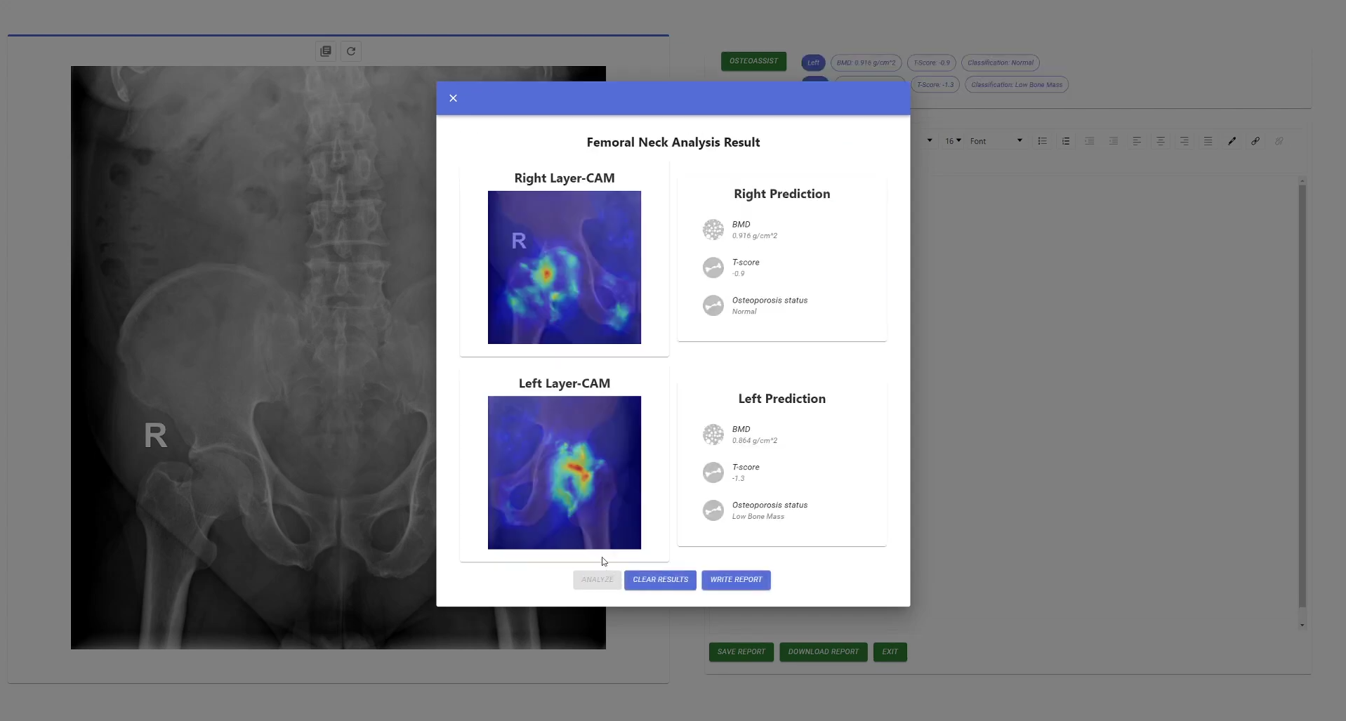
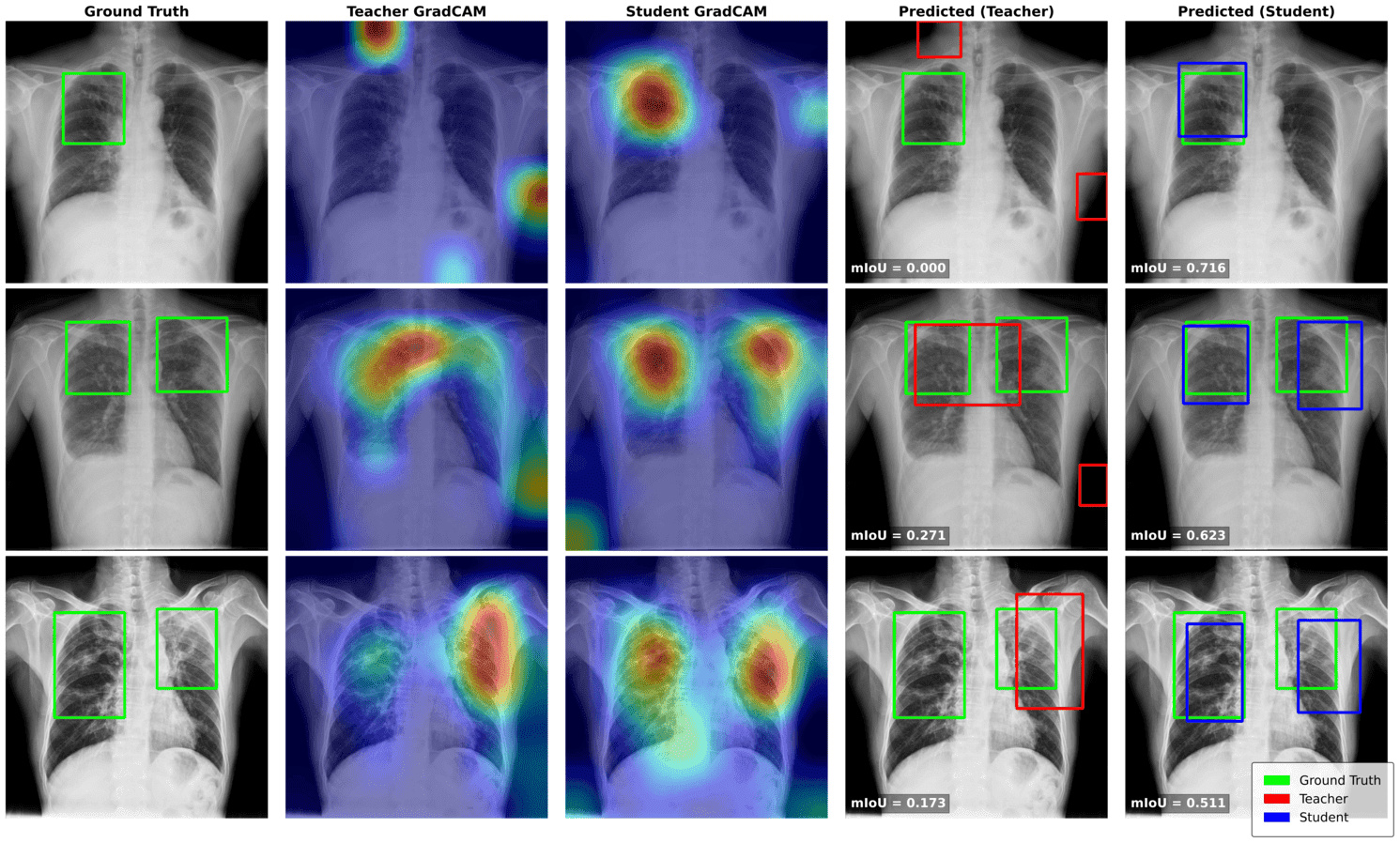
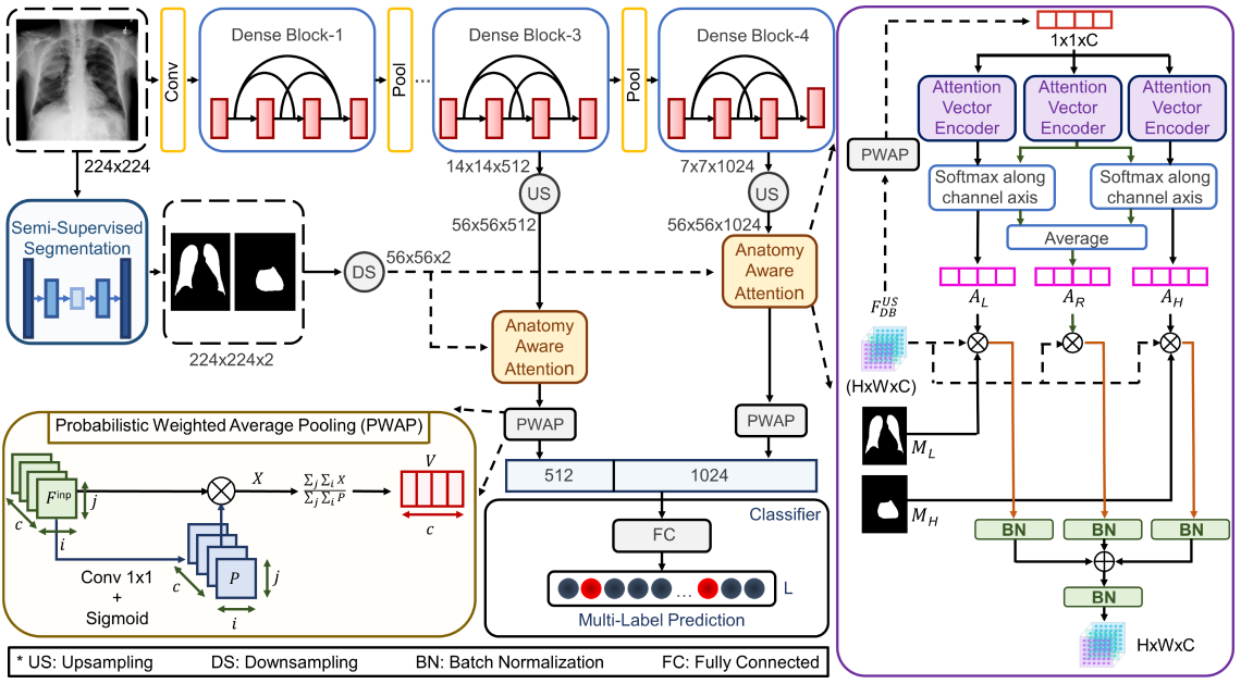
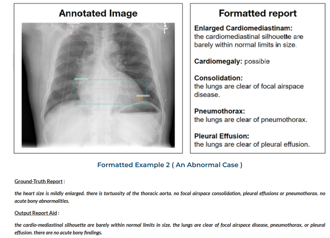
In our lab, we develop AI-driven systems that assist radiologists by automatically analyzing chest X-rays and generating structured report drafts. Our research focuses on combining image understanding with medical language generation to translate visual abnormalities detected in X-rays into clinically meaningful descriptions. By integrating disease localization, semantic understanding, and language modeling, we aim to produce reports that remain grounded in visual evidence.


2024 - 2025

AI in Screening of Orthopaedic Conditions

Screening of Orthopaedic Conditions

Osteoporosis is a significant global health concern, particularly in LMICs like Bangladesh, where diagnostic limitations hinder early detection. With 43.2% of females and 30.3% of males at risk, osteoporosis remains undiagnosed until fractures occur, leading to disability and reduced quality of life. On the other hand, osteoarthritis is the most common degenerative joint disease that can lead to chronic pain, stiffness, and loss of function, and it is recognized by the World Health Organization as a significant contributor to years lived with disability worldwide.

We focus on common orthopaedic conditions such as osteoporosis and osteoarthritis, building tools that can learn from routine X-rays and relevant clinical information to identify disease, estimate bone health, and assess severity in a more objective way. Instead of depending only on specialized tests that may be expensive or difficult to access, we aim to unlock more value from imaging that is already widely used in everyday care. By combining medical imaging, patient data, and modern deep learning methods, we create practical decision-support technologies that can help clinicians screen patients earlier, prioritize follow-up, and better understand disease progression.


2024 - 2025

Trustworthy TB Detection

TB Detection

A common problem with artificial intelligence in healthcare is that models can sometimes cheat by focusing on irrelevant parts of an image instead of actual signs of disease. Our research addresses this issue by developing a more disciplined way for models to learn how to identify tuberculosis. We used a framework that encourages the AI to ignore distractions and instead focus on the specific areas of the lungs where abnormalities occur. This breakthrough is important because it makes the technology more trustworthy for doctors to use in real-world situations.


2022

Thoracic disease detection from X-ray Images

Thoracic Disease Detection from X-ray

Thoracic diseases such as pneumonia, cardiomegaly, and pleural abnormalities are commonly evaluated using chest X-rays, one of the most widely used imaging tools in clinical practice. Although deep learning models have shown strong potential for automated detection of these conditions, their performance often declines when applied across different hospitals, imaging devices, and patient populations. Variations in imaging style, anatomical differences, and shifts between adult and pediatric cases can significantly affect model reliability.

In our lab, we investigate AI methods that improve the robustness and clinical relevance of chest X-ray analysis. Our research focuses on incorporating anatomical knowledge, developing learning strategies that reduce sensitivity to domain differences, and enabling knowledge transfer across diverse patient groups. By guiding models toward clinically meaningful structures and improving their ability to generalize across datasets, we aim to build chest X-ray diagnostic systems that remain dependable in real-world healthcare environments.

Publications